- Book an appointment:
+90 212 417 21 21 -


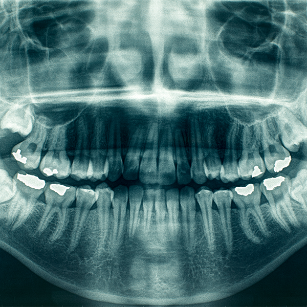
What is a Dental X-ray?
Dental X-rays, also known as Radiographs, are images that dentists use to see a patient's oral health and condition. They are applied to see the mouth and tooth structure. By using a Dental X-ray, the dentist can see decays, gaps, and the position of the wisdom tooth. In general, it is as important as a dental cleaning.
Why Are Dental X-Rays Applied?
Dentists usually apply Dental X-rays to their patients once a year. If the patient has a current treatment or a condition that the dentist needs to follow, several x-rays may be requested.
Factors such as the patient's age, current oral health, caries, or gum problems may affect the frequency of X-rays.
Dentists request X-rays from new patients who have never had X-rays in the past.
Children's teeth need to be followed during their growth process, so they may need more X-rays than adults. It is especially important to prevent situations where milk teeth and adult teeth overlap.
Are Dental X-Rays Risky?
Since Dental X-rays contain very low levels of radiation, they are not dangerous for children or adults. A protective bib is placed on the patient's chest during the X-ray. Children and pregnant women usually use this bib.
X-rays are risky for pregnant women and cannot be applied. Women who have the potential to become pregnant should also inform their dentists about the situation.
Is Preparation Required for a Dental X-Ray?
No special preparation is required. Oral cleaning before the X-ray appointment may help employees.
X-rays are taken in the dentist's office, with the patient sitting in a chair and wearing a protective bib on their chest. The device is placed near the patient's head and it records images inside of the mouth. Some dentists have a separate room for X-rays.
What are the Types of Dental X-Rays?
There are several different types of X-rays, although Intraoral X-rays are the most common. Intraoral X-rays are also divided into types themselves.
Bitewing: The patient bites off a piece of paper so that the dentist can see the condition of the veneers. It is preferred to see the spaces between the teeth.
Occlusal technique: It is applied to see the condition of the patient's lower and upper teeth. The jaw should be in a closed position. The floor of the mouth and palate can also be checked with this technique.
Panoramic technique: X-rays are taken while the machine rotates around the patient's head. The wisdom tooth and implants usually are checked with this method.
Preapical: Used to see a tooth entirely from root to crown.
In addition, the Extraoral technique can be used in some special cases.
A competent person in the field accompanies the patient at all stages of the X-ray. It is only necessary to leave the patient alone while the images are taken.
After the extraction is over, the dentist examines the images. If a problem is found, treatment is planned with the patient.